Model 502
Feature
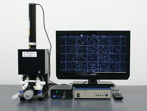
When an external voltage is applied to a colloidal dispersion, charged particles move toward one of the electrodes. The zeta potential of the particles is determined by measuring their migration velocity under a microscope. Since the particles can be observed directly, this is a reliable method that avoids the "black box" issue.
Because observations are limited to particles suspended at the stationary plane, the results are unaffected by electroosmotic flow.
The system produces high-contrast images, enabling the measurement of ultrafine particles as small as 20 nm.
There are two methods for determining migration velocity:
1. Stopwatch method
The time taken for individual particles to travel a specific distance is measured using a stopwatch.
2. Image analysis method (optional)
The electrophoretic process is recorded, and image analysis is used to calculate the migration velocity of individual particles.
Specification
| Method |
Electrophoretic mobility measurement using the stopwatch method
|
| Range | ±100 mV |
| Available size range | 0.02 ~ 50 μm(depending on particle) |
| Concentation | less than 0.001 wt%(depending on particle) |
| Supply voltage |
0 ~ 160 V(stop watch method) |
| Cell | Quartz glass |
| Sample volume | 3 ~ 10 mL |
| Light source | 2 mW He-Ne Laser |
| Microscope | ×280 |
| Camera | CCD Monochrome TV camera |
| Display | 19 inch TV |
| Voltage | 100 V、50/60 Hz |
| Option |
Image analysis to determine electrophoretic mobility N-type non-aqueous electrophoresis cell unit |
Case Studies
Zeta potential of alumina as a function of pH
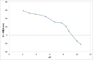
Sample: A 0.1 M KCl dispersion of high-purity α-alumina (AKP-30).
Measurements of the zeta potential across a pH range of 2 to 11 yielded an isoelectric point of approximately 9.3, which is in good agreement with the literature value of 9.0–9.5.
● Application
Powders, metal oxides, ultrafine metal particles, pigments, emulsions, polymer latices, photographic emulsions, viscous materials, clay, carbon black, carbon nanotubes, pulp slurries, cellulose nanofibers, surfactants, flocculants, biological particles, blood cells, microorganisms, colloidal particles, etc.